
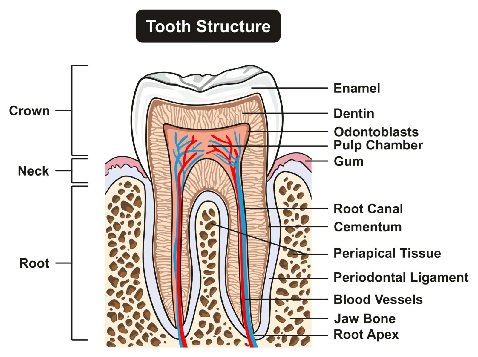
Structure of Tooth Diagram
3D MODEL Tooth eruption There is a broad range of normal times for teeth to push through the gum tissue (erupt) into the mouth. For primary teeth, the central incisors are the first teeth to erupt, occurring at about 6 months of age. These are followed by the lateral incisors, first primary molars, canines, and, finally, second primary molars.
What's Inside Your Teeth? Acorn Dentistry For Kids
Introduction This e-Anatomy module contains fifty four illustrations dedicated to the anatomy of the teeth. These fully annotated anatomical illustrations are presented as a comprehensive atlas of the dental anatomy specifically designed for students in dentistry and medicine, residents and healthcare professionals. Material and methods
The Purpose of Teeth Dr. Kevin Sands
Introduction Toothache, and other dental problems, are common presenting complaints in both primary and secondary care. 1 Having a basic understanding of the dentition (or teeth) is important for all healthcare professionals.

Human Tooth Diagram Stock Illustration Download Image Now iStock
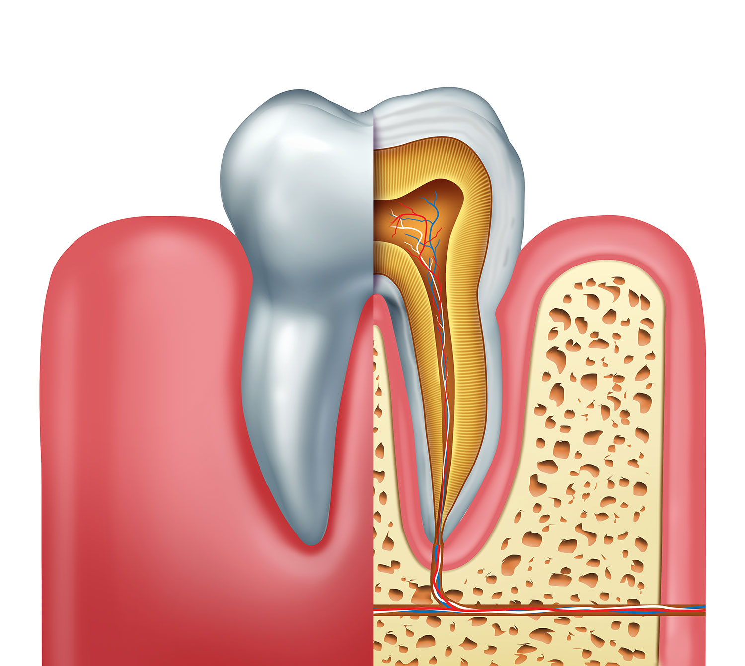
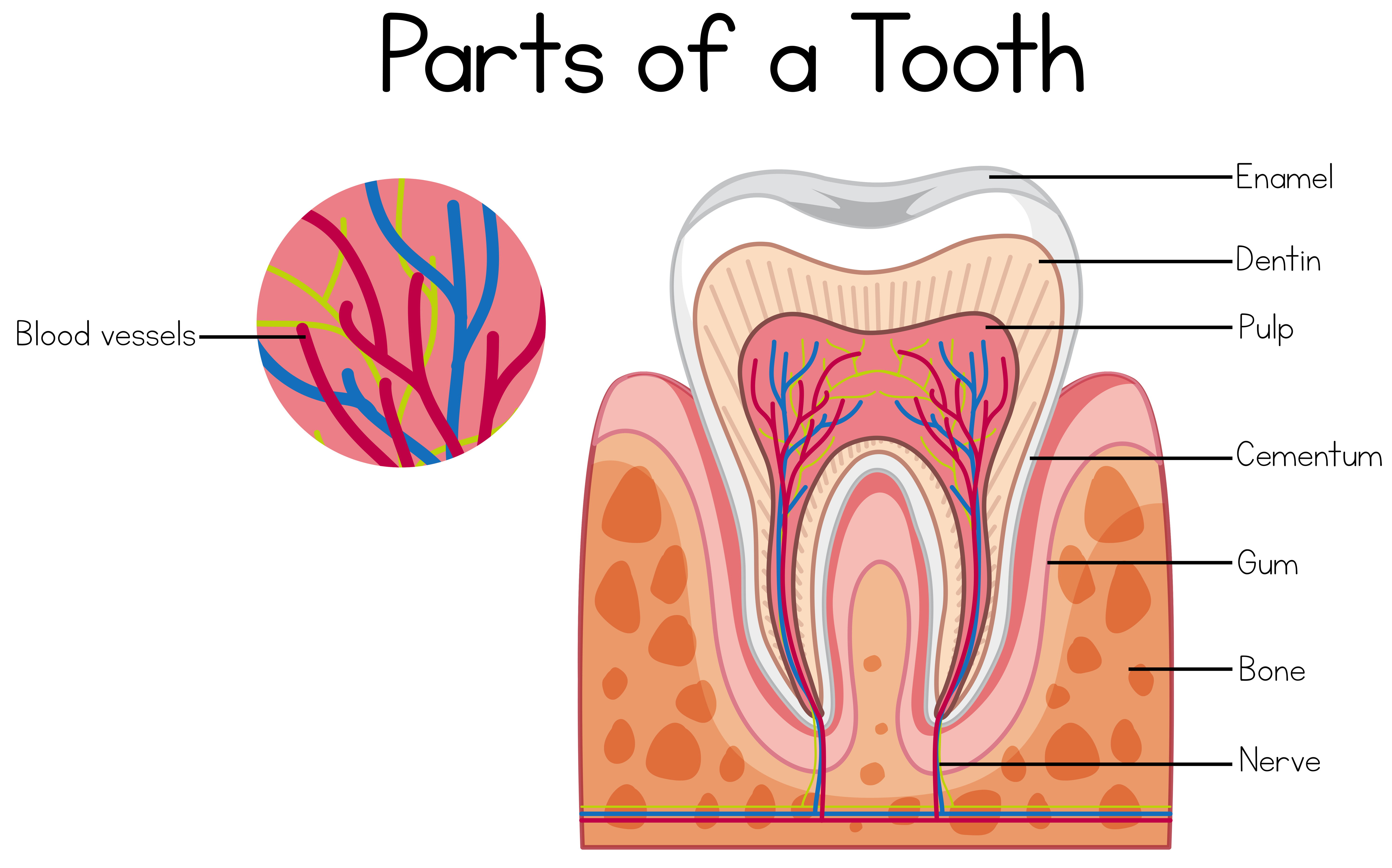
Teeth anatomy Tooth structure Arteries and veins Nerves Sources Related articles + Show all Teeth names and numbering There are thirty-two teeth in total in the oral cavity of an adult dentition. One half, or sixteen, are embedded in the maxilla, while the lower half are situated within the mandible.

Anatomy of the Teeth Anatomical Chart Anatomy Models and Anatomical
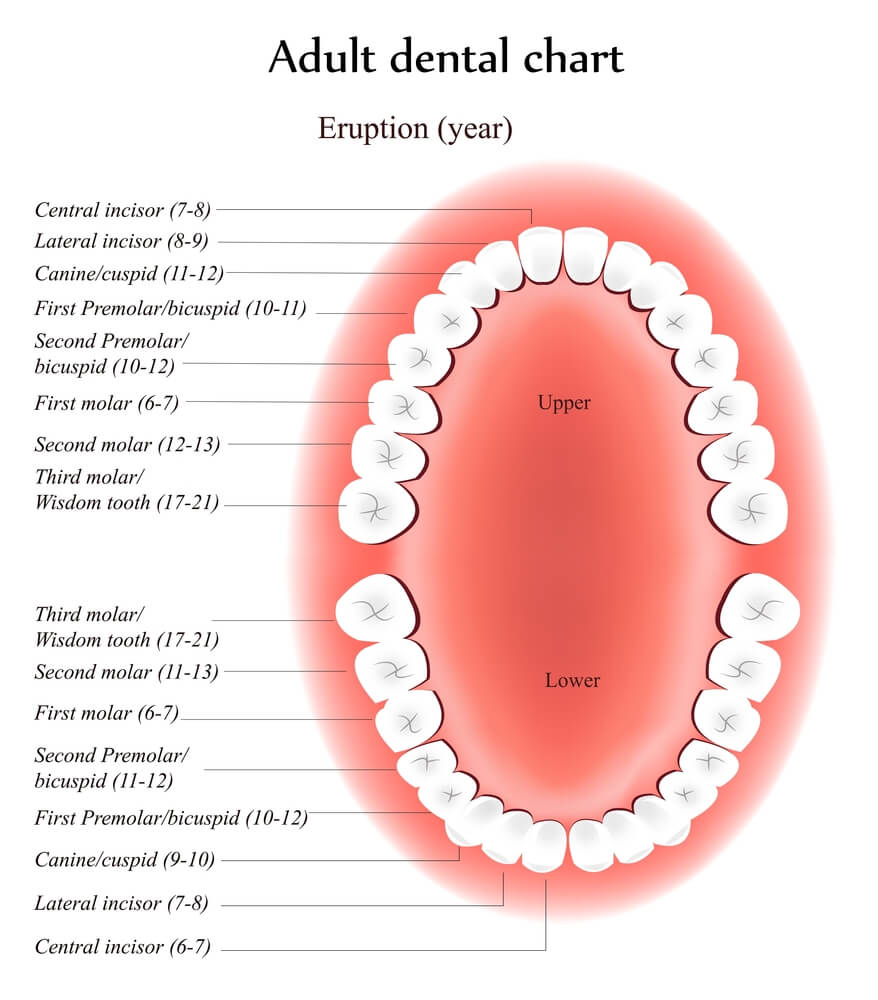
Tooth diagram Tooth conditions Symptoms of a tooth condition Health tips Types of teeth Most people start off adulthood with 32 teeth, not including the wisdom teeth. There are four.
The Anatomy and Structure of a Tooth Tuxedo Dental Group
Teeth diagram: numbering and incisal surfaces Each incisor has five surfaces, each one named according to the anatomical structure that it faces: Labial surface - faces the lips. Lingual surface - faces of the mandibular incisors face the tongue. The corresponding maxillary incisors have a palatal surface instead of a lingual one that faces the.
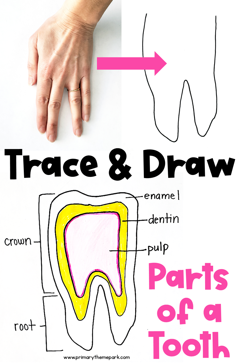
Parts of a Tooth Diagram Primary Theme Park
Learn about the types of teeth in a fast and efficient way using our interactive tooth identification quizzes and labeled diagrams. This leaves up to eight adult teeth in each quadrant and separates the opposing pairs within the same alveolar bone as well as their counterparts in the opposing jaw. Each quadrant contains: a medial incisor

anatomy of the teeth chart
The "control" in these two diagrams consists of beams of coronal dentin prepared with diamond slicing wheels, and with average surface roughness (Ra) of less than 0.2 μm.. Cracks on the tooth's surface must undergo a combination of growth towards the DEJ (i.e. longitudinally along the rods) and about the occlusal surface (i.e.
Parts of a tooth diagram 614137 Vector Art at Vecteezy
Human teeth function to mechanically break down items of food by cutting and crushing them in preparation for swallowing and digesting. As such, they are considered part of the human digestive system. [1] Humans have four types of teeth: incisors, canines, premolars, and molars, which each have a specific function.

Diagram of a human tooth [150]. Download Scientific Diagram
This diagram helps us learn the names of each tooth, the corresponding number, and their location. While brushing or searching for cavities, knowing about teeth numbers can help you identify and explain issues to your dentist.

Tooth Number Chart to Identify Primary Teeth Eruption Charts
tooth, any of the hard, resistant structures occurring on the jaws and in or around the mouth and pharynx areas of vertebrates. Teeth are used for catching and masticating food, for defense, and for other specialized purposes. The teeth of vertebrates represent the modified descendants of bony dermal (skin) plates that armoured ancestral fishes.
The Anatomy of Your Teeth
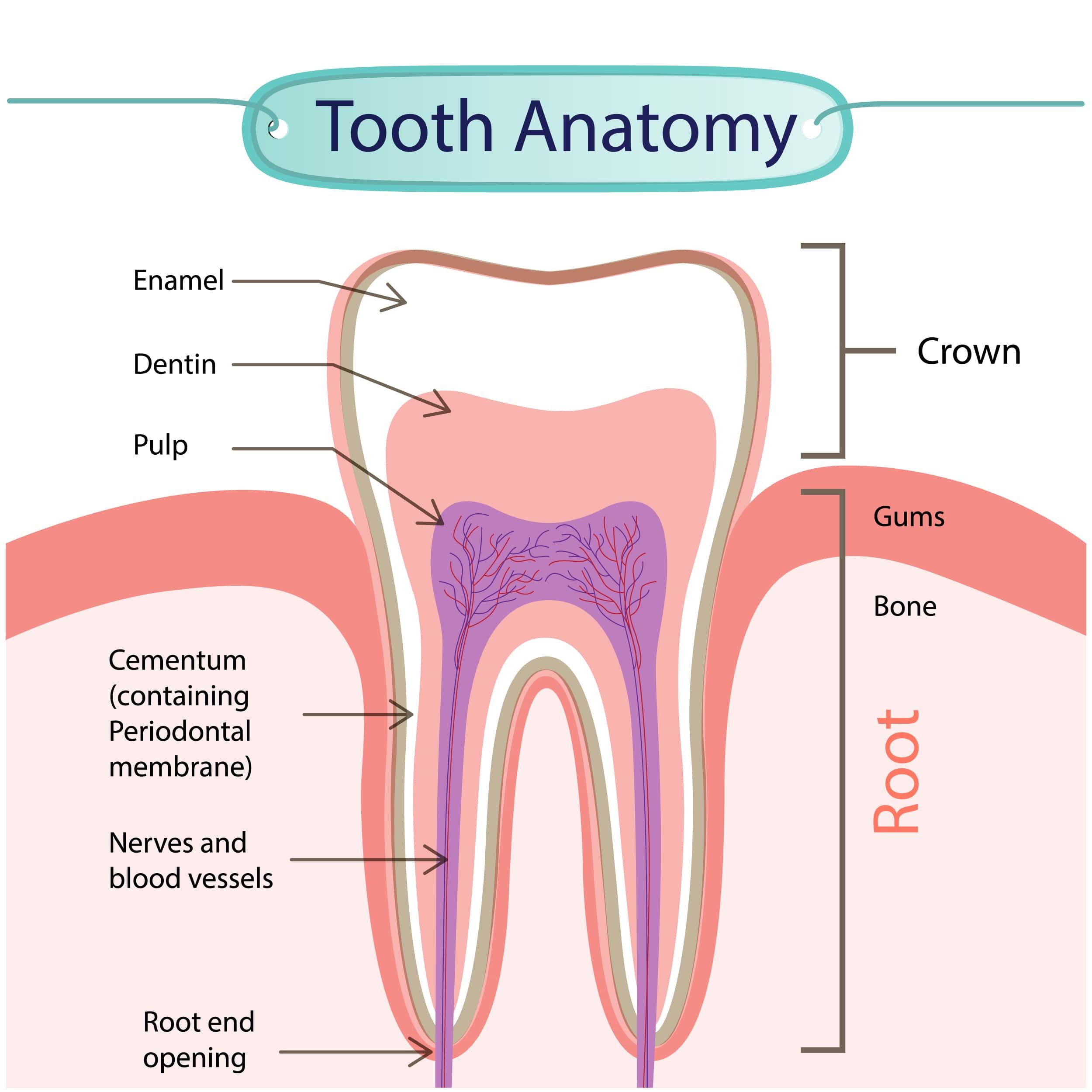
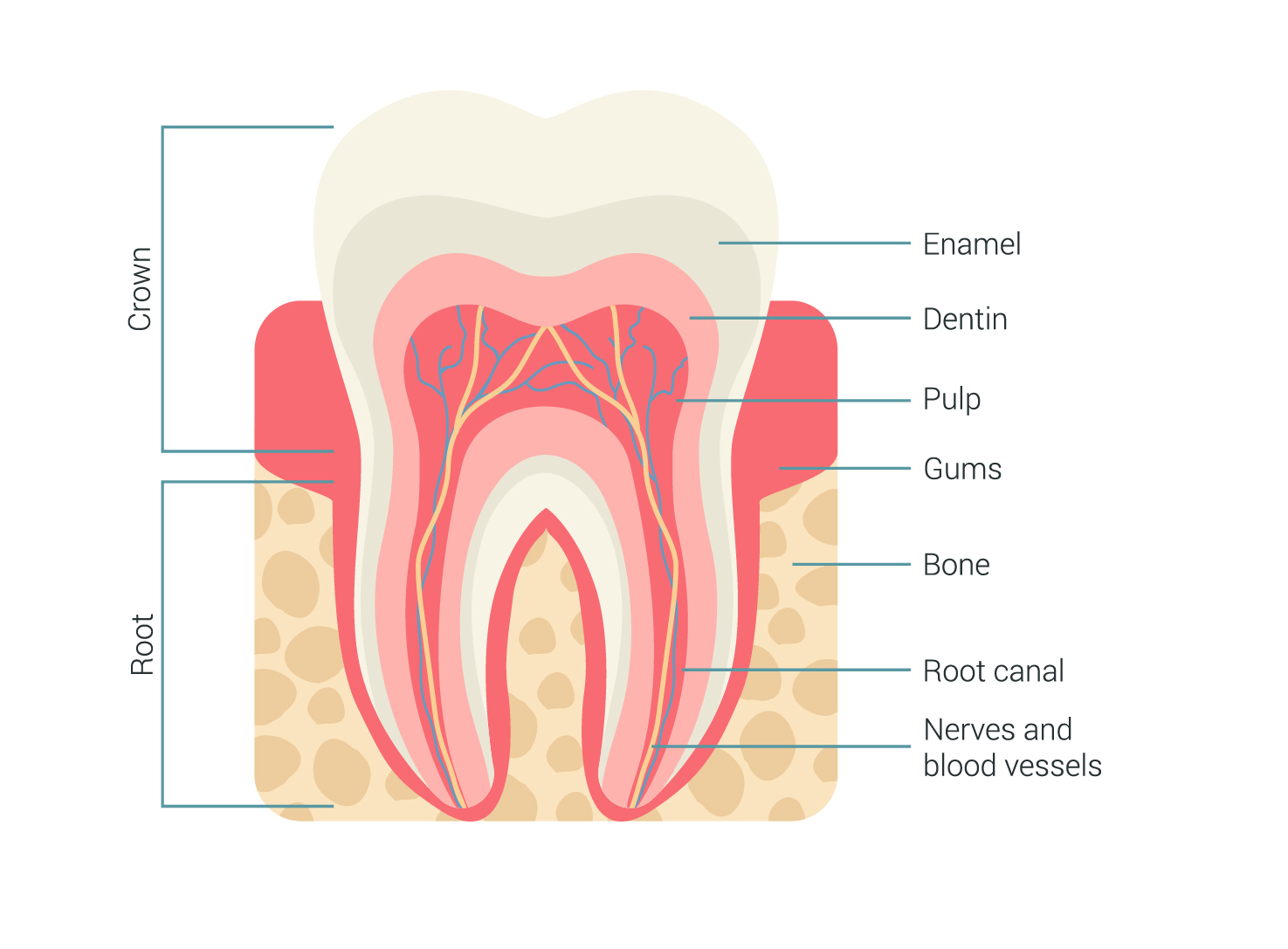
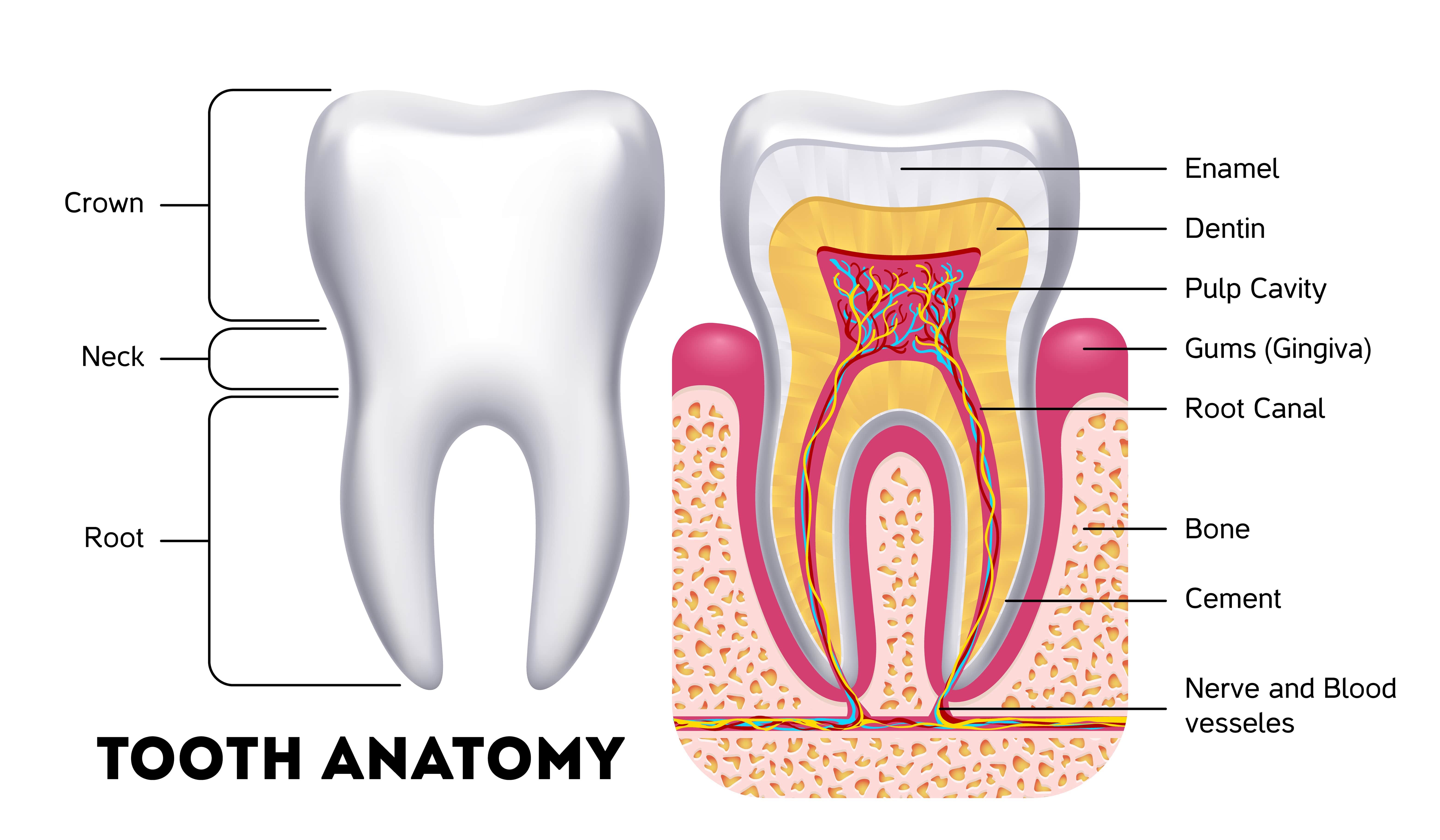
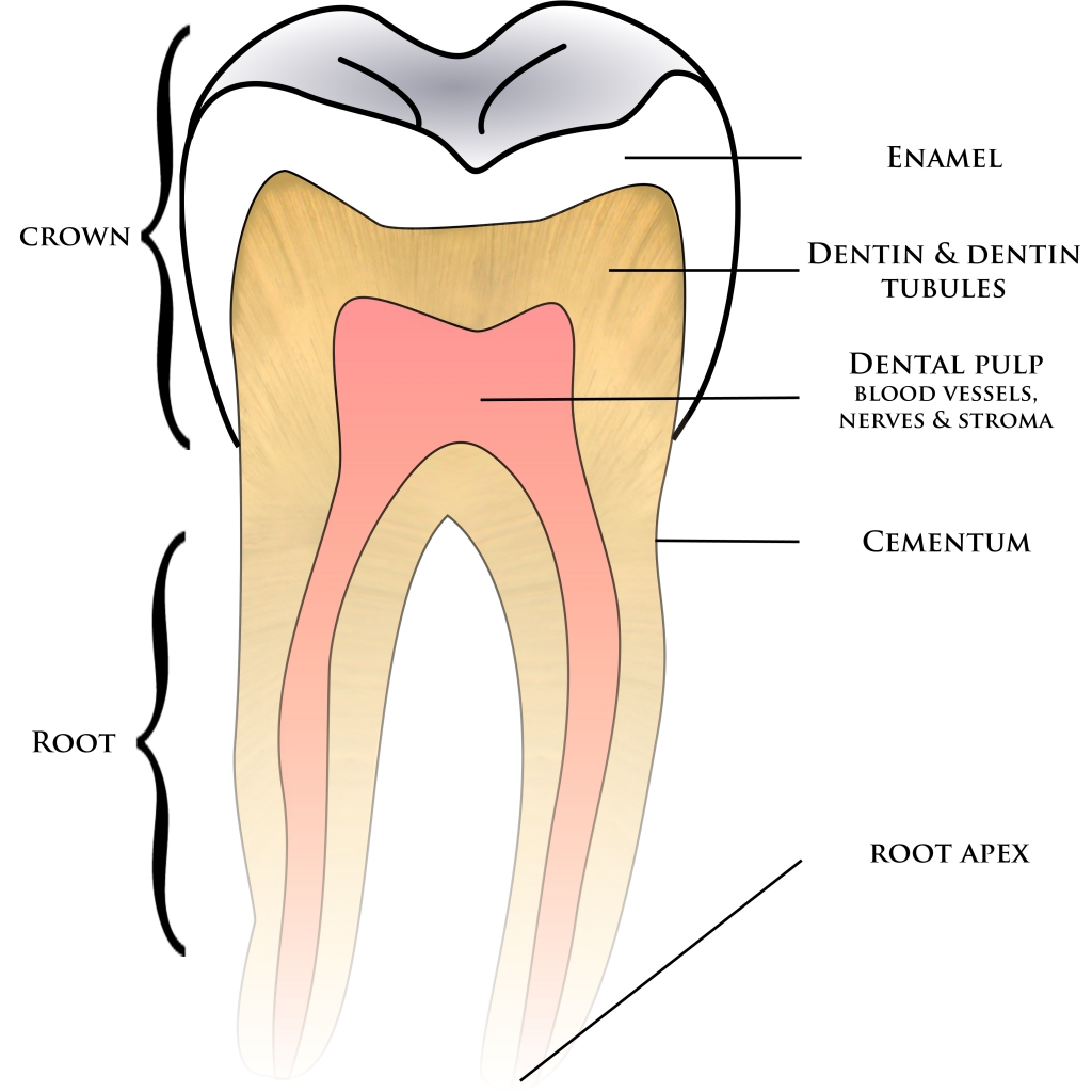
The crown refers to the part of a human tooth that is visible to us. The enamel, dentin, cementum, pulp, root, periodontal ligaments, etc., are important parts of the tooth structure. Bodytomy provides labeled human tooth diagrams to help you understand the human tooth anatomy.
Tooth Anatomy Milford Family Dentistry
Canines Canines are the sharp, pointed teeth that sit next to the incisors and look like fangs. Dentists also call them cuspids or eyeteeth. Canines are the longest of all the teeth, and people.
The Anatomy Of A Tooth In Four Parts Arc Dental
To record changes to your dental health, dentists use a chart with a diagram of your teeth. The teeth are numbered according to the Universal Numbering System adopted by the American Dental Association. The diagram is drawn as if you're looking at your dentist with your mouth wide open. The top teeth are numbered from right to left.
Dental and Oral Anatomy Star Dental Assisting School
Diagram of the Tooth Numbering System (viewed as if looking into the mouth) Buccal (Facial) Surface Occlusal Surface Incisal Surface Right Left Maxillary Arch (Upper Jaw) Mandibular Arch (Lower Jaw) Adult Dentition = Permanent teeth 1-32 Child Dentition =Primary teeth A-T Wisdom Teeth =1, 16, 17, and 32 Central Incisor Lateral Incisor Cuspid.

6 Procedures to Repair a Broken or Cracked Tooth
Dental anatomy Dental anatomy is a field of anatomy dedicated to the study of human tooth structures. The development, appearance, and classification of teeth fall within its purview. (The function of teeth as they contact one another falls elsewhere, under dental occlusion .)